Accurate, Less Time, More Success: Jaipur Doctors Used 3D Printed Model to Practise Surgery
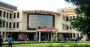
Doctors at the Sawai Man Singh hospital in Jaipur performed a delicate surgery by first working out its details on a 3D printed model. It helped them save time, avoid complications and save the patient's life.
In Jaipur, doctors at the Sawai Man Singh hospital used 3D printing to create a model of a craniovertebral junction (the head and neck) to practise surgery before attempting it on a living human.
The surgery was conducted on March 22 on a young patient who was suffering from craniovertebral junction (CVJ) anomaly for five years. Diagnosed in February, the condition made it difficult for him to move his neck, along with weakness in his limbs. A surgery was mandatory, but it ran a risk of severely disabling the patient.

Claiming to be the first in the country to use this technique, Dr Rashim Kataria, assistant professor of neurosurgery at the hospital, and his team, successfully completed the surgery.
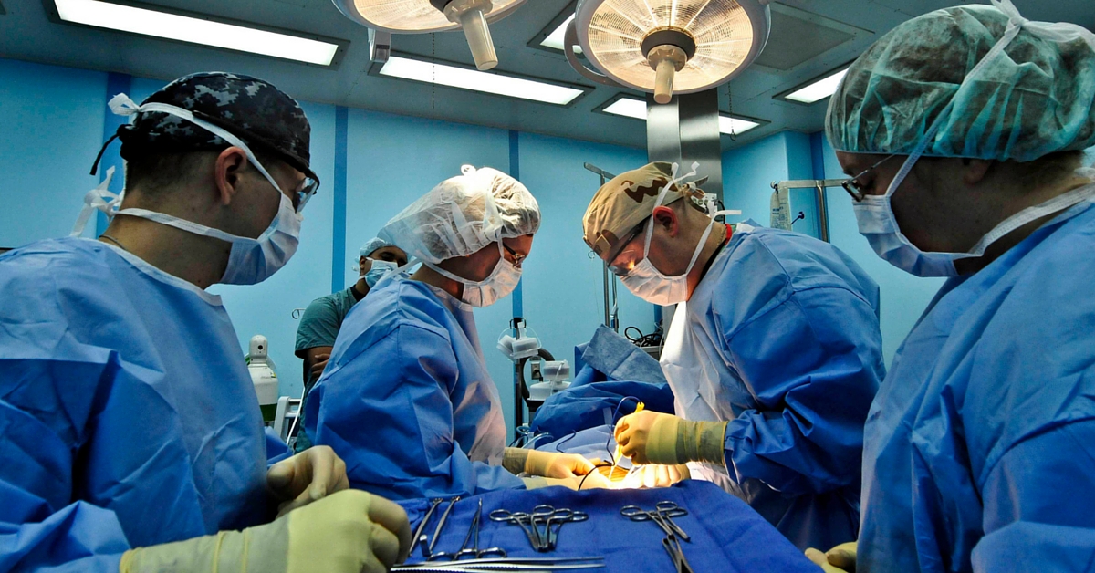
Source: Wikimedia Commons
Explaining how the surgery was conducted, he said to Times of India, “We practise surgery on the model of CV Junction, which is exactly the same CV Junction of the patient. It allows us to know about the nerves and other anatomy of the head and neck junction. When we conducted the surgery, we already had in mind the exact location of nerves and CV Junction, which was to be operated.”
The benefits of this technique are many. It minimised chances of error and improved accuracy by figuring out exactly where to operate. The doctors could confidently perform the operation, without much blood loss, in much lesser time.
The patient, who now recovered successfully, was discharged from the hospital on Saturday, April 2.
If this is just the start, then 3D printing can be used as a way to figure out surgical details for a lot more surgeries that are complicated. The technique is already under trial at the Massachusetts Institute of Technology (MIT) in the US. The scientists there devised a 3D printer that could assess a patient’s heart and print a model in a few hours. The device includes software that can analyse MRI scans and create clear, precise scans. What’s more, the 3D model can also be useful when the doctors have to explain conditions in medical terms. The visual representation helps patients and their families to understand what the doctor is saying in a much clearer context.
Like this story? Or have something to share? Write to us: [email protected], or connect with us on Facebook and Twitter (@thebetterindia).
This story made me
-
 97
97 -
 121
121 -
 89
89 -
 167
167
Tell Us More

We bring stories straight from the heart of India, to inspire millions and create a wave of impact. Our positive movement is growing bigger everyday, and we would love for you to join it.
Please contribute whatever you can, every little penny helps our team in bringing you more stories that support dreams and spread hope.