Indian Scientist Develops Prototype Of A Radiation-Free Breast Cancer Detection Machine
A team of scientists led by Prof. Srirang Manohar is working on a very promising prototype to improve breast cancer detection rates. The technology uses a new form of imaging technique and is also radiation-free. Read on to know more about how this imaging machine can change the way breast cancer is diagnosed today.

Breast cancer is the most common form of cancer in women worldwide.
It is especially on the rise in developing countries where usually the diagnosis occurs at later stages.
Worldwide, yearly about 450,000 women die from the consequences of breast cancer. Current imaging modalities are not optimal in discriminating benign (non-cancerous) from malignant (cancerous) tissue. Owing to less-optimal imaging detection techniques, there are numerous cases of breast cancer that lead to late diagnosis.


Prof. Srirang Manohar & Team Is Making A Change
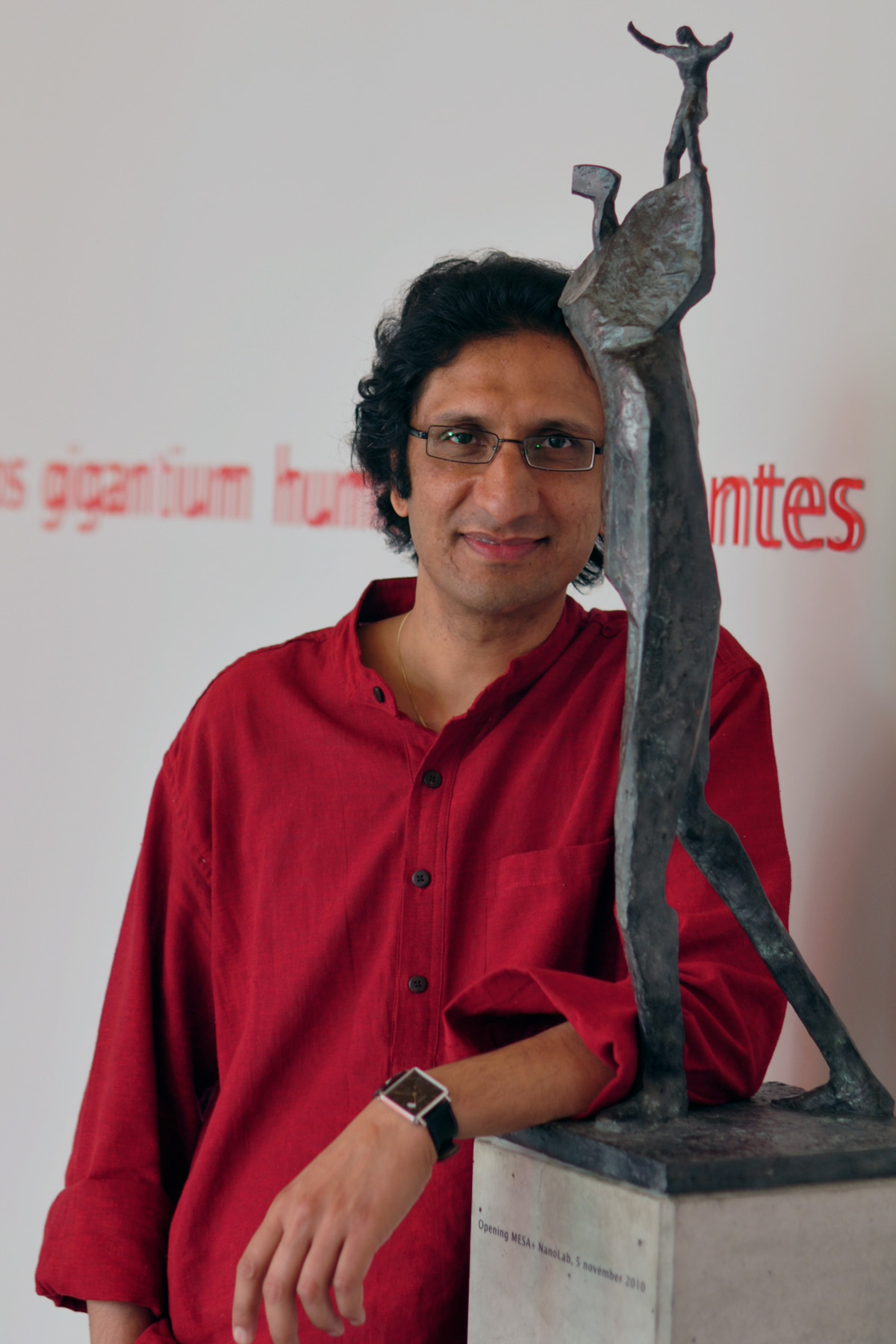
Meet Srirang Manohar, an Associate Professor, in the Faculty of Science and Technology’s Biomedical Photonic Imaging Group (BMPI) at the University of Twente in the Netherlands. He is one of the pioneers in the research and development of a sophisticated breast cancer diagnostic instrument, the TWENTE PHOTOACOUSTIC MAMMOSCOPE (PAM). His group investigates the use of light for medical purposes. Their final aim is to develop optical and hybrid optical-acoustical technologies for medical diagnosis, in particular in the fields of oncology and wound healing.
When asked to describe his current area of research, Siring said,
“I work broadly in Biomedical Optics, which is the study and application of light interactions with biological tissue. Specifically my interests lie in photoacoustic imaging which I apply for the detection and diagnosis of breast cancer and rheumatoid arthritis”.
The University of Twente’s PHOTOACOUSTIC MAMMOSCOPE (PAM)
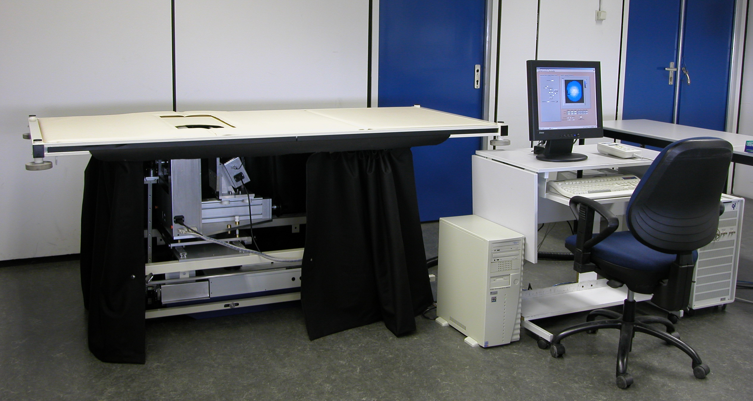
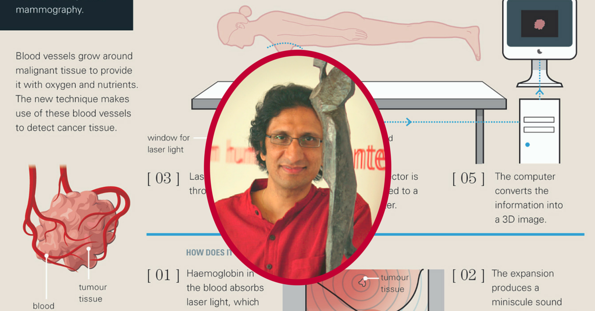
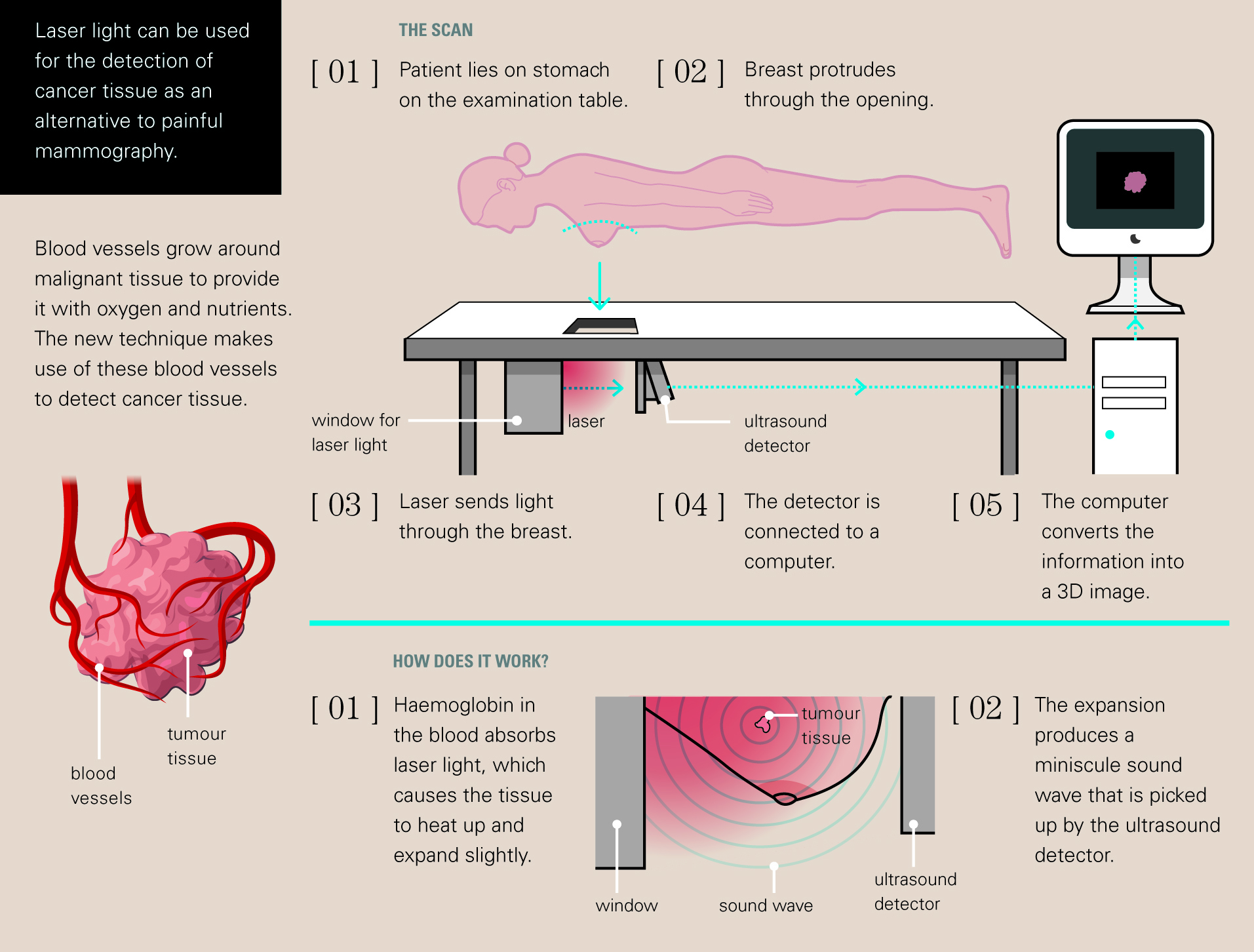
A team of researchers led by Associate Professor Srirang Manohar Biomedical Imaging Group at the University of Twente, in the Netherlands have developed a prototype of a new imaging tool that may one day help to detect breast cancer early, when it is most treatable. If effective, the new device called a photoacoustic mammoscope (PAM) would represent an entirely new way of imaging the breast and detecting cancer. Instead of X-rays, which are used in traditional mammography, the Photoacoustic Mammoscope uses a combination of infrared light and ultrasound to create a 3-D image of the breast.
Recently, a large clinical study has been started by Srirang and his group with the Medisch Spectrum Twente in Oldenzaal using PAM.
The Twente Photoacoustic Mammoscope (PAM), which was first tested in 2007, has since been upgraded with several new features which are expected to improve the resolution as well as add the capability to image using several different wavelengths of light at once, which in turn is expected to improve detectability.
Photoacoustics
In the search for improved imaging modalities for detection and diagnosis of breast cancer, the ability to differentiate between benign cysts and malignant lesions is of great importance. Photoacoustic (PA) imaging is a relatively new imaging modality that has potential for visualizing breast malignancies
To create a photoacoustic image, pulses of laser light are shone onto the tissue being scanned. The light scatters inside tissue and gets selectively absorbed by tumors. This is because tumors are associated with high density of blood vessels, and hemoglobin in blood absorbs light strongly. The absorbed light is converted to heat leading to a temperature rise, – just a few thousandths of a degree – that is perfectly safe but not enough to cause tumor to expand abruptly due to thermoelastic expansion. This leads to the emission of sound waves in the ultrasonic range. An array of sensors placed on the skin picks up these waves, and a computer then uses a process of triangulation to turn the ultrasonic signals into a two- or three-dimensional image of what lies beneath. It is an imaging modality that relies on listening to the “sound” generated by “light.”
Manohar and his colleagues added that if the instrument were commercialized, it would likely cost less than MRI and X-ray mammography and probably not much more than an ultrasound machine.
When asked to describe his ‘eureka moment’, Srirang had this to say,
“ My high point so far was when I presented the first clinical results of the instrument that I had developed to detect breast cancer at a conference in the United States, in 2007. The hall was jam-packed and a there was a buzz of anticipation when I made my presentation, just a triumphant 15 minute long, which condensed experiences of 5 years of hard work, inspiration and teamwork, plus some depressing moments too”.
Visualizing the malignancy-associated increased haemoglobin concentration might significantly improve early diagnosis of breast cancer. This where the Photoacoustic Mammoscope developed could help in early detection of breast cancer.
About Prof. Srirang Manohar
Srirang Manohar was born in Bengaluru and did his early schooling at St. Joseph’s Boys High School, followed by MES College. He then went on to complete his B.E from the R.V. College of Engineering, Bengaluru. Srirang then graduated with an M.S and PhD from the Department of Instrumentation (now renamed as the Department of Instrumentation and Applied Physics), Indian Institute of Science, Bengaluru. In 2001 Srirang went to the University of Twente, the Netherlands as a Post-Doctoral Fellow and took up research in the area of Biomedical Optics. He is the recipient of several awards, among which is the prestigious VENI Scholarship by the Dutch organization for Scientific Research. On a lighter vein, Srirang loves South Indian delicacies, especially the Masala Dosa.
All Photographs Courtesy: Prof. Srirang Manohar.
This story made me
 97
97 121
121 89
89 167
167
Tell Us More
We bring stories straight from the heart of India, to inspire millions and create a wave of impact. Our positive movement is growing bigger everyday, and we would love for you to join it.
Please contribute whatever you can, every little penny helps our team in bringing you more stories that support dreams and spread hope.